Bone Cross Section Diagram - Cross Section Of Bone Png Image Transparent Png Free Download On Seekpng / Explaned distal and proximal epiphysis.. 71558774, anatomy, artery, biomedical illustration, bone marrow tissue, bones, cancellous bone, cavity, colour image, compact, connective tissue, connective tissues, dense bone, dental filling, diaphysis, health, healthcare and medicine. Descubre ilustraciones de la más alta calidad de human bone cross section diagram of femur showing osteon veins marrow. Diagram with articular cartilage, marrow, medullary cavity and periosteum. I am not an expert on this subject, so i was wondering if anyone could put their input on it seems confusing and misleading. Healthy tooth diagram isolated on white background vector.
Explaned distal and proximal epiphysis. Explaned distal and proximal epiphysis. The centroidal locations of common cross sections are well documented, so it is typically not necessary to calculate the location with the equations above. They are similar to the topographic profiles that you created in the topographic maps chapter, but they also show the rock types and geologic structures. A cross section diagram is if you would take a knife and cut through one side of a diagram to see the inside and outside in one picture.
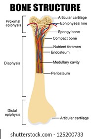
Bone Cross Section High Res Stock Images Shutterstock from image.shutterstock.com In this short video i use blender 2.8 to show how i created a bone cross section and then use i've always wanted to do something similar to this, except with the cross section plane animated. Diagram with articular cartilage, marrow, spongy bone, medullary cavity, endosteum, diaphysis, and periosteum. Cross section diagrams are used a lot by architects and engineers to show what a building or machine might look like before it's built. As shown in figure 2. Download 130+ royalty free bone cross section vector images. Vector illustration scheme of bone cross section. Cross section through middle metacarpal bones of vector. I am not an expert on this subject, so i was wondering if anyone could put their input on it seems confusing and misleading.
Explaned distal and proximal epiphysis. Detailed and high textured 4k normal,disp,diffuse. Descubre ilustraciones de la más alta calidad de human bone cross section diagram of femur showing osteon veins marrow. Vector illustration scheme of bone cross section. The centroidal locations of common cross sections are well documented, so it is typically not necessary to calculate the location with the equations above.
Procedure Part 1 1 Locate These Structures And Label Chegg Com from media.cheggcdn.com Vector illustration scheme of bone cross section. Jump to navigation jump to search. A cross section of a human long bone. They are similar to the topographic profiles that you created in the topographic maps chapter, but they also show the rock types and geologic structures. Learn vocabulary, terms and more with flashcards, games and other study tools. Diagram with articular cartilage, marrow, spongy bone, medullary cavity, endosteum, diaphysis, and periosteum. Cross section diagrams are used a lot by architects and engineers to show what a building or machine might look like before it's built. This is a short tutorial using blender 2.8 that shows how to create a bone cross section and using images to create the textures.hope you enjoy and please.
Descubre ilustraciones de la más alta calidad de human bone cross section diagram of femur showing osteon veins marrow.
The centroidal locations of common cross sections are well documented, so it is typically not necessary to calculate the location with the equations above. Diagram with articular cartilage, marrow, spongy bone, medullary cavity, endosteum, diaphysis, and periosteum. Learn vocabulary, terms and more with flashcards, games and other study tools. Vector illustration scheme of bone cross section. Explaned distal and proximal epiphysis. Each system contains haversian canals surrounded by concentric lamellae of bone tissue 48. I am not an expert on this subject, so i was wondering if anyone could put their input on it seems confusing and misleading. Diagram with articular cartilage, marrow, medullary cavity and periosteum. Download 130+ royalty free bone cross section vector images. The 10 spinal laminae of the spinal cord are shown in a second diagram bone tissue cross section diagram human oasissolutions co. They build the entire picture, improve your understanding, consolidate the information and facilitate recall. As shown in figure 2. Explaned distal and proximal epiphysis.
They build the entire picture, improve your understanding, consolidate the information and facilitate recall. Vector illustration scheme of bone cross section. Explaned distal and proximal epiphysis. Detailed and high textured 4k normal,disp,diffuse. Descubre ilustraciones de la más alta calidad de human bone cross section diagram of femur showing osteon veins marrow.
Bone Anatomy Ask A Biologist from askabiologist.asu.edu Health, bones, one object, vein, human skeleton, artery, cavity, skeletal system, nerve, compact, human bone, human tissue, human nervous system, marrow, spongy bone, porous, connective tissue, spongy, human artery, cancellous bone, diaphysis. A cross section of a human long bone. Cross section through middle metacarpal bones of vector. Download 130+ royalty free bone cross section vector images. Cross section of a human bone. From wikimedia commons, the free media repository. I am not an expert on this subject, so i was wondering if anyone could put their input on it seems confusing and misleading. The centroidal locations of common cross sections are well documented, so it is typically not necessary to calculate the location with the equations above.
A cross section diagram is if you would take a knife and cut through one side of a diagram to see the inside and outside in one picture.
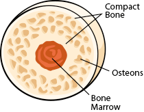
71558774, anatomy, artery, biomedical illustration, bone marrow tissue, bones, cancellous bone, cavity, colour image, compact, connective tissue, connective tissues, dense bone, dental filling, diaphysis, health, healthcare and medicine. Descubre ilustraciones de la más alta calidad de human bone cross section diagram of femur showing osteon veins marrow. Vector illustration scheme of bone cross section. From wikimedia commons, the free media repository. Compact bone is the outer layer and the spongy bone forms the inner layer. Spinal cord spinal column anatomy information myvmc. They are similar to the topographic profiles that you created in the topographic maps chapter, but they also show the rock types and geologic structures. A cross section diagram is if you would take a knife and cut through one side of a diagram to see the inside and outside in one picture. Health, bones, one object, vein, human skeleton, artery, cavity, skeletal system, nerve, compact, human bone, human tissue, human nervous system, marrow, spongy bone, porous, connective tissue, spongy, human artery, cancellous bone, diaphysis. This is a short tutorial using blender 2.8 that shows how to create a bone cross section and using images to create the textures.hope you enjoy and please. Explaned distal and proximal epiphysis. Vector illustration scheme of bone cross section. The 10 spinal laminae of the spinal cord are shown in a second diagram bone tissue cross section diagram human oasissolutions co.
Jump to navigation jump to search bone cross section. Each system contains haversian canals surrounded by concentric lamellae of bone tissue 48.


0 Komentar